
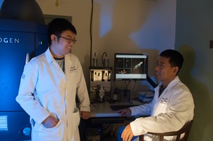
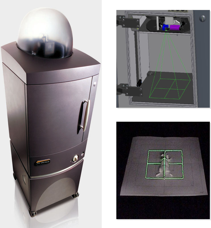
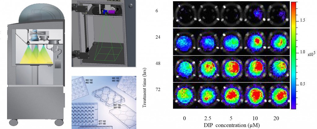
The laboratory has a PerkinElmer IVIS® Spectrum imaging system which has brought physics and biology together for the practice of real-time in vivo imaging. It is one of the most versatile and advanced in vivo imaging system available on the market today. This IVIS Imaging System includes a sensitive CCD camera, a dark imaging chamber to minimize incident light, and specialized software to quantify and analyze the results.
 For advanced fluorescence imaging, the IVIS Spectrum has the capability to use either trans-illumination (from the bottom) or epi-illumination (from the top) to illuminate in vivo fluorescent sources. 3D diffuse fluorescence tomography can be performed to determine source localization and concentration using the combination of structured light and trans illumination fluorescent images. The instrument is equipped with 10 narrow band excitation filters (30nm bandwidth) and 18 narrow band emission filters (20nm bandwidth) that assist in significantly reducing autofluorescence by the spectral scanning of filters and the use of spectral unmixing algorithms. In addition, the spectral unmixing tools allow the researcher to separate signals from multiple fluorescent reporters within the same animal.
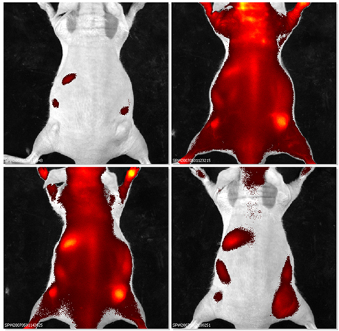
The IVIS Imaging Systems are currently being used for genomics, biotechnology, and pharmaceutical product research and development. Data gathered from animal models based on the real-time in vivo imaging technology allow researchers to better understand the mechanisms of diseases and drug effects.
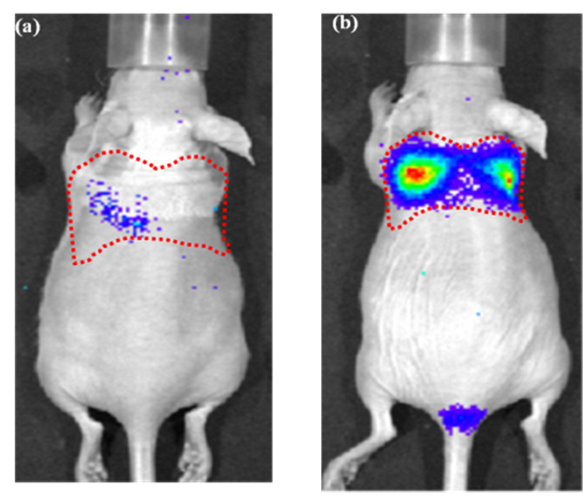
HC11 mammary epithelial cells transfected with a luciferase construct of the beta-casein gene promoter (p-344/-1betac-Lux). The promoter activity was imaged optically in real time following lactogenic induction. The imaging signal intensity was closely correlated with that measured using a luminometer following protein extraction and consistent with the messenger RNA (mRNA) level of the endogenous beta -casein gene.