Advances in neuroimaging technology have provided a wealth of information regarding brain development and injury in children with congenital heart disease. In conjunction with clinical research, preclinical imaging studies using the translational porcine model will aid in acquiring a full picture of impaired brain development and neurological injury associated with congenital heart disease.
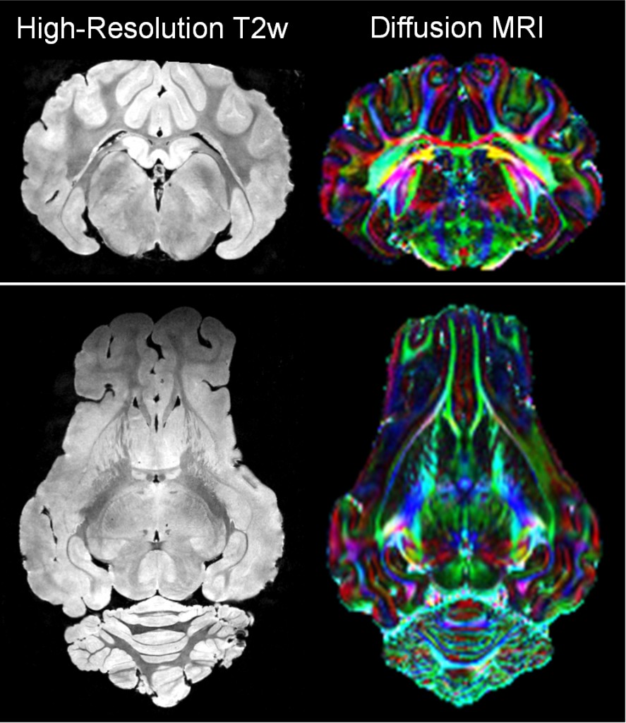
High-resolution brain MRI of Yorkshire piglet model of congenital heart disease. T2w MRI and diffusion MRI demonstrate contrast of piglet brain anatomy and white matter tracks for visualizing brain injury in congenital heart disease.
References
Stinnett GR, Lin S, Korotcov AV, Korotcova L, Morton PD, Ramachandra SD, Pham A, Kumar S, Agematsu K, Zurakowski D, Wang PC, Jonas RA, Ishibashi N. Microstructural Alterations and Oligodendrocyte Dysmaturation in White Matter After Cardiopulmonary Bypass in a Juvenile Porcine Model. J Am Heart Assoc. 2017 Aug 15;6(8):e005997.doi: 10.1161/JAHA.117.005997. PMID 28862938PMC5586442