imaging-research-mrs

Research Highlight: Biomedical Imaging

Molecular MR Imaging and Spectroscopy on Brain Injury and Cancer

Molecular MR imaging and spectroscopy provide non-invasive detection of metabolites in the brain tissue and tumor to avoid invasive examinations. This study applies and CEST imaging and NMR spectroscopy to investigate the metabolic profiles in neonatal hypoxia and immunotoxin therapy of glioblastoma.

CEST MRI Imaging
Molecular CEST MRI of U87-EGFRvIII tumor xenografts.
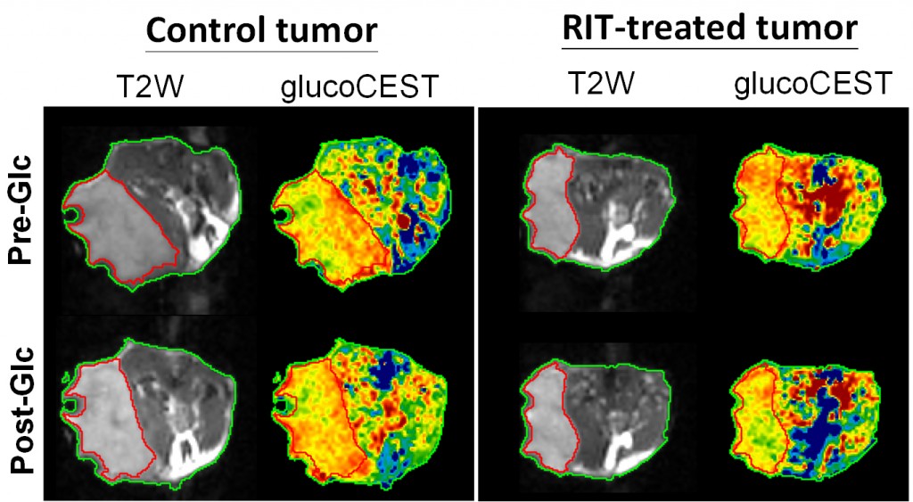
CEST imaging detects a distinct pattern of glucose uptake between a tumor treated by DT390-BiscFv806 recombinant immunotoxin treatment (RIT) and a tumor treated by PBS vehicle. The results suggest that the RIT could significantly inhibit the growth of an established U87-EGFRvIII glioblastoma tumor xenograft.
1H-MRS of hippocampal metabolites in the in vivo and in situ mouse brain.
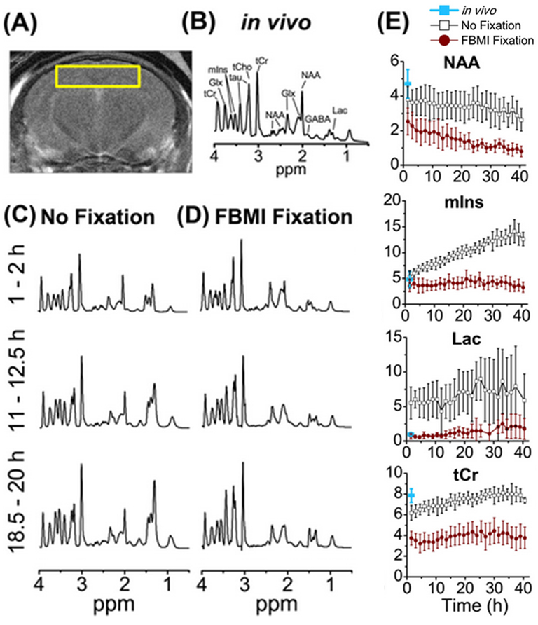
(A) T2w MRI of a mouse brain labelled with a voxel of interest on hippocampus for 1H-MRS of (B) in vivo, (C) no fixation, and (D) focused beam microwave irradiation (FBMI) fixation. (E) Comparison of hippocampal metabolite concentrations between the data acquired by in vivo (under isoflurane anesthesia) and in situ (no fixation and FBMI fixation) 1H-MRS.
References
  • Meng J, Liu YY, Gao SY, Lin S, Gu XB, Pomper MG, Wang PC, Shan L.
    A Bivalent Recombinant Immunotoxin with High Potency against Tumors with EGFR and EGFRvIII Expression.
    Cancer Biol Ther. Oct 2015. PMID 26467217 PMC4847807
  • Hsu CH, Lin S, Ho AC, Johnson TD, Wang PC, Scafidi J, Tu TW.
    Comparison of in vivo and in situ detection of hippocampal metabolites in mouse brain using 1H-MRS.

    NMR Biomed. 2020 Nov 30:e4451. doi: 10.1002/nbm.4451. Epub ahead of print. PMID: 33258202 PMID 33258202