The laboratory has two state-of-the-art NMR machines: a Bruker Avance III 7 Tesla (300 MHz), 21cm horizontal bore MRI/MRS system and a Bruker Avance 9.4 Tesla (400 MHz), 89 mm vertical bore MRI/MRS system. Both MRI/MRS systems are capable of performing MRI as well as spectroscopy studies. The NMR laboratory has acquisition workstations, a storage server and several workstations for spectroscopy data and image processing.
Bruker 7T MRI
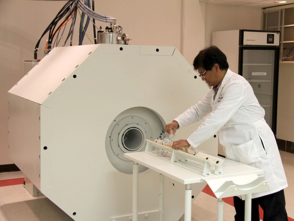
Bruker 7 Tesla (300MHz) MRI/MRS
210mm horizontal bore Magnex magnet, 112mm clear space
RF Coils: Volume, surface, and custom-made coils
AVANCE III Console, Paravision 6 (PV6) software
Detectable nuclei: 1H
Bruker 9.4T MRI
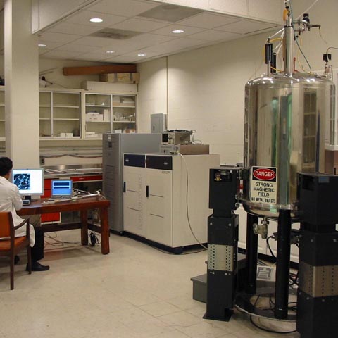
Bruker 9.4 Tesla (400MHz) MRI/MRS
89mm vetical bore Oxford magnet
RF Coils: Volume, surface, and custom-made coils
AVANCE 400 Console, Paravision 5.1 (PV5) software
Detectable nuclei: 1H, 19F, 13C, 31P
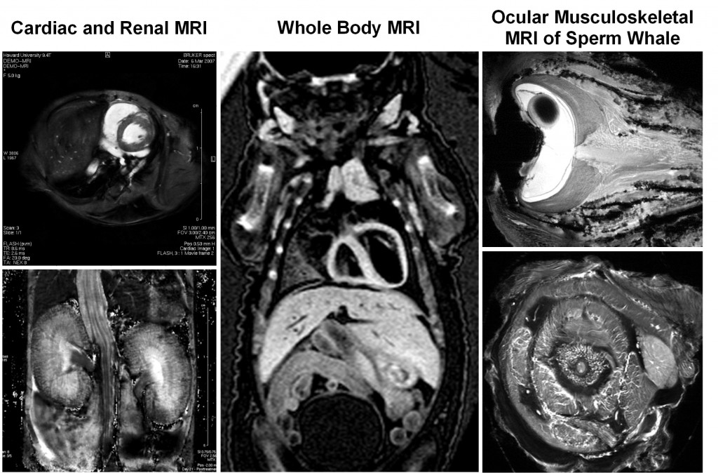
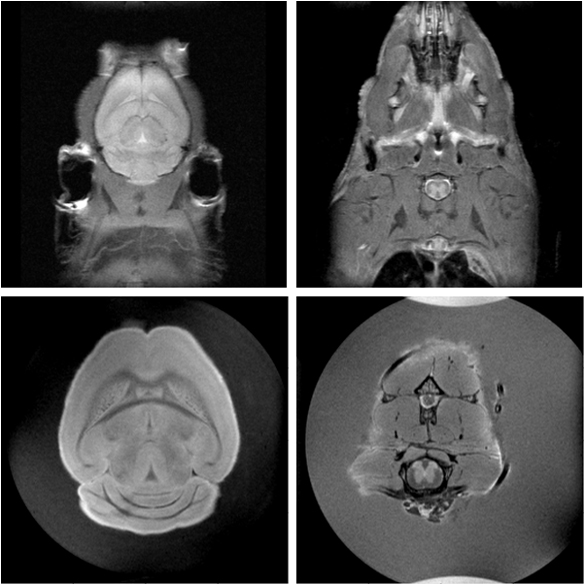
Sample MRI Images
Example MRI studies for animal models of cardiac, renal, gastroenterological and musculoskeletal diseases and biology.
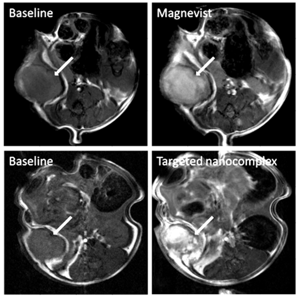
A mouse tumor (MDA-MB-231 solid tumor xenograft marked with arrow) visualized using Dynamic Contrast Enhanced MRI with Magnevist (upper row) and a targeted (Transferrin-Liposome-Magnevist) nanocomplex (lower row). The targeted nanocomplex exhibited a stronger and heterogeneous signal enhancement.
In vivo (upper row) and ex vivo (lower row) normal mouse brain images visualized using a high resolution T2 weighted fast spin-echo MRI sequence. This sequence is designed to detect Alzheimer's pathology (amyloid plaques) in the brains and spinal cords of transgenic mice, which become detectable by MRI at 6-8 months of disease progression.